
Page 58 - 89Zr-Immuno-PET:Towards a Clinical Tool to Guide Antibody-based Therapy in Cancer
P. 58
Chapter 3
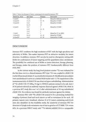
Table 5. Tumor involvement of tumor containing levels missed by immuno-PET
Patient no., level
1, level II 3, level III 5, level III
6, level II
7, level I 14, level IV 15, level II
Size (mm) %Tumor
7 x 11 25
9 x 15 50 - 75 4x 4 10
8 x 15 10
5x 7 5
6x 8 50 - 75 5x 9 5
5x 7 5
8 x 12 10 - 25 5x 5 25 - 50
REMARKS
Missed by MRI/detected by CT Missed by CT/MRI
Missed by CT/MRI
Missed by CT/MRI
Missed by CT (no MRI) Missed by CT/MRI Detected by CT/MRI
DISCUSSION
Immuno-PET combines the high resolution of PET with the high specificity and selectivity of MAbs. This makes immuno-PET an attractive modality for tumor detection. In addition, immuno-PET can also be used in a therapeutic setting with MAbs for confirmation of tumor targeting and for quantitative dose calculations. The possibility for combined use of MAbs in tumor detection, therapy planning, and therapy, makes the position of immuno-PET fundamentally different from FDG-PET.
In the current study, the long-lived positron emitter 89Zr was evaluated for the first time ever in a clinical immuno-PET trial. 89Zr was coupled to cMAb U36 via the bifunctional chelate N-succinyldesferrioxamine B. Modification procedures were standardized to arrive at a chelate/MAb molar ratio <2. No impairment of the immunoreactivity of cMAb U36 was observed upon radiolabeling. Administration of 89Zr-cMAb U36 (75 MBq, 10 mg) to HNSCC patients seemed to be safe. Just 2 of 20 patients showed an antibody response directed against cMAb U36, while in a previous RIT study this was 5 of 12 after administration of 50 mg radiolabeled cMAb U36. No evidence was found for antibody reactions against the chelate.
Immuno-PET with 89Zr-cMAb U36 seemed to be a promising method for imaging of primary head and neck tumors as well as metastases in the neck. All primary tumors were visualized, whereas 18 of 25 tumor-containing neck levels were also identified. In this feasibility study, the sensitivity of immuno-PET for detection of lymph node metastases was at least as good as of CT/MRI: 72% versus 60%. In a previous SPECT study with 99mTc-labeled mMAb U36 in a comparable
56
