
Page 55 - 89Zr-Immuno-PET:Towards a Clinical Tool to Guide Antibody-based Therapy in Cancer
P. 55
Immuno-PET with 89Zr-cMab U36 in head and neck cancer
3
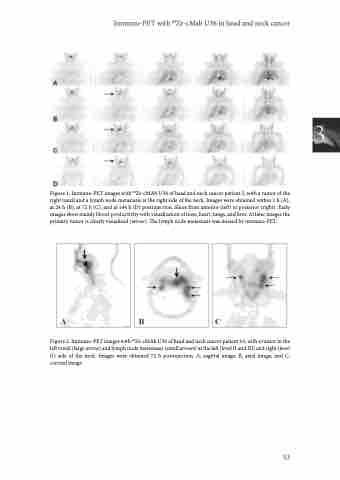
Figure 1. Immuno-PET images with 89Zr-cMAb U36 of head and neck cancer patient 5, with a tumor of the right tonsil and a lymph node metastasis at the right side of the neck. Images were obtained within 1 h (A), at 24 h (B), at 72 h (C), and at 144 h (D) postinjection. Slices from anterior (left) to posterior (right). Early images show mainly blood-pool activity with visualization of nose, heart, lungs, and liver. At later images the primary tumor is clearly visualized (arrow). The lymph node metastasis was missed by immuno-PET.
Figure 2. Immuno-PET images with 89Zr-cMAb U36 of head and neck cancer patient 16, with a tumor in the left tonsil (large arrow) and lymph node metastases (small arrows) at the left (level II and III) and right (level II) side of the neck. Images were obtained 72 h postinjection. A, sagittal image; B, axial image, and C, coronal image.
53
