
Page 186 - 89Zr-Immuno-PET:Towards a Clinical Tool to Guide Antibody-based Therapy in Cancer
P. 186
Chapter 9
RESULTS
Patlak Linearization
Per patient, Patlak linearization was conducted for each tissue (kidney, liver, lung, spleen) (Fig.2). An r-value of >0.9 was obtained for 71/80 linear fits for 89Zr- antiPSMA and 89Zr-antiHER2 (3 late time points available). Therefore, we assumed that data for 89Zr-antiCD20 and 89Zr-EGFR were consistent with the assumptions under the Patlak method. For these datasets, the r-value for the linear fit could not be obtained (only 2 late time points available). For 89Zr-antiHER2 an r-value <0.9 was obtained for 1/10 fits for kidney uptake, 2/10 for liver, 4/10 for lung and 2/10 for spleen. The mean percentage difference between fitted and measured tissue activity concentrations per time point for all fits through 3 time points with r >0.9 was ±4%, except for 89Zr-antiPSMA in the kidney (Supplemental Table 2).
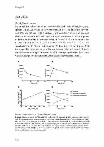
Figure 2 Transfer constants for 89Zr-antiHER2 in the kidney
Example for one patient in the 89Zr-antiHER2 study, with (A) measured activity concentrations in serum and (B) measured activity concentrations in the kidney. Patlak linearization (C) to determine the offset (VT) and slope (Ki) of the linear fit to the last three time points (same data). (D) Reversible (red line) and irreversible (blue line) contributions to the total measured signal (black line). No target expression has been reported for HER2 in the normal kidney. Therefore, we hypothesize that the total signal consists of non- specific uptake. After 100h p.i., total uptake predominantly consists of irreversible non-specific uptake due to 89Zr-residualization after mAb degradation.
184
