
Page 126 - 89Zr-Immuno-PET:Towards a Clinical Tool to Guide Antibody-based Therapy in Cancer
P. 126
Chapter 6
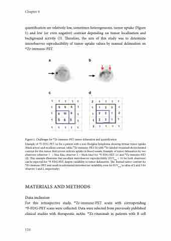
quantification are relatively low, sometimes heterogeneous, tumor uptake (Figure 1) and low (or even negative) contrast depending on tumor localization and background activity (5). Therefore, the aim of this study was to determine interobserver reproducibility of tumor uptake values by manual delineation on 89Zr-immuno-PET.
Figure 1. Challenges for 89Zr-immuno-PET tumor delineation and quantification
Example of 18F-FDG-PET (a) for a patient with a non-Hodgkin lymphoma showing intense tumor uptake (black arrow) and excellent contrast, while 89Zr-immuno-PET (b) with 89Zr-labeled-rituximab shows limited contrast for this tumor. Red arrows indicate uptake in blood vessels. Example of tumor delineation by two observers (observer 1 = blue line, observer 2 = black line) for 18F-FDG-PET (c) and 89Zr-immuno-PET (d). This example illustrates that excellent interobserver reproducibility (SUVmax = 10 for both observers) can be expected for 18F-FDG-PET, despite variability in tumor delineation. The limited tumor contrast for 89Zr-immuno-PET may result in substantial interobserver variability, even for SUVmax (a value of 2 and 3 for observer 1 and 2, respectively).
MATERIALS AND METHODS
Data inclusion
For this retrospective study, 89Zr-immuno-PET scans with corresponding 18F-FDG-PET scans were collected. Data were selected from previously published clinical studies with therapeutic mAbs: 89Zr-rituximab in patients with B cell
124
