
Page 90 - Advanced concepts in orbital wall fractures
P. 90
88
Chapter 5
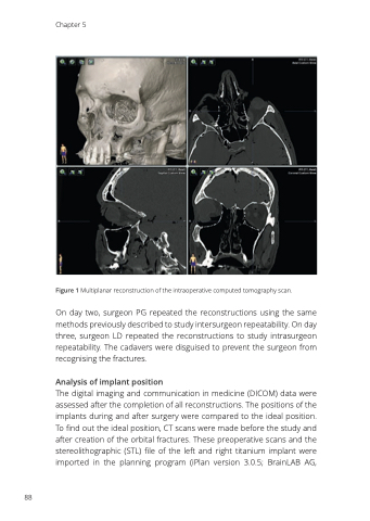
Figure 1 Multiplanar reconstruction of the intraoperative computed tomography scan.
On day two, surgeon PG repeated the reconstructions using the same methods previously described to study intersurgeon repeatability. On day three, surgeon LD repeated the reconstructions to study intrasurgeon repeatability. The cadavers were disguised to prevent the surgeon from recognising the fractures.
Analysis of implant position
The digital imaging and communication in medicine (DICOM) data were assessed after the completion of all reconstructions. The positions of the implants during and after surgery were compared to the ideal position. To find out the ideal position, CT scans were made before the study and after creation of the orbital fractures. These preoperative scans and the stereolithographic (STL) file of the left and right titanium implant were imported in the planning program (iPlan version 3.0.5; BrainLAB AG,
