
Page 56 - Advanced concepts in orbital wall fractures
P. 56
54
Chapter 3
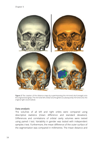
Figure 3 The creation of the distance maps by superimposing the mirrored skull (orange) onto the original skull (grey). The mirrored left orbital volume (green) is subsequently mirrored onto the original right volume (blue).
Data analysis
The volumes of all left and right orbits were compared using descriptive statistics (mean difference and standard deviation). Differences and correlations of orbital cavity volumes were tested using paired t test. Variability in gender was tested with independent samples t test. Furthermore, the mean difference of the outer surface of the segmentation was compared in millimetres. The mean distance and
