
Page 84 - Bladder Dysfunction in the Context of the Bladder-Brain Connection - Ilse Groenendijk.pdf
P. 84
82
Chapter 4
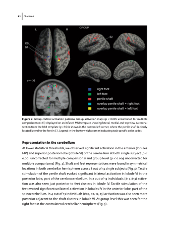
Figure 2. Group cortical activation patterns. Group activation maps (p < 0.005 uncorrected for multiple comparisons; n=13) displayed on an inflated MNI template showing lateral, medial and top view. A coronal section from the MNI template (y=-36) is shown in the bottom-left corner, where the penile shaft is clearly located lateral to the feet in S1. Legend in the bottom-right corner indicating task specific color codes.
Representation in the cerebellum
At lower statistical thresholds, we observed significant activation in the anterior (lobules I-IV) and superior posterior lobe (lobule VI) of the cerebellum at both single subject (p < 0.001 uncorrected for multiple comparisons) and group level (p < 0.005 uncorrected for multiple comparisons) (Fig. 3). Shaft and feet representations were found in symmetrical locations in both cerebellar hemispheres across 8 out of 13 single subjects (Fig. 3). Tactile stimulation of the penile shaft evoked significant bilateral activation in lobule VI in the posterior lobe, part of the cerebrocerebellum. In 2 out of 13 individuals (#11, #13) activa- tion was also seen just posterior to feet clusters in lobule IV. Tactile stimulation of the feet evoked significant unilateral activation in lobules IV in the anterior lobe, part of the spinocerebellum. In 4 out of 13 individuals (#04, 07, 12, 13) activation was also seen more posterior adjacent to the shaft clusters in lobule VI. At group level this was seen for the right foot in the contralateral cerebellar hemisphere (Fig. 3).
