
Page 74 - Postoperative Intra-Abdominal Adhesions- New insights in prevention and consequences
P. 74
Chapter 4
Results
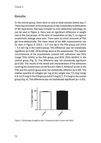
In the control group, there were no sick or dead animals before day 7. There was one death in the study group on day 3 secondary to dehiscence of the laparotomy. Necropsy showed no intra-abdominal pathology. As can be seen in Figure 1, there was no significant difference in weight loss in the two groups. At the time of reoperation on day 7, no signs of anastomotic leakage were seen. There were no visual remnants of PVA gel intra-abdominally. The mean result of the ABP measurements can be seen in Figure 2: 155.0 – 6.7 mm Hg in the PVA group and 173.0 – 7.5 mm Hg in the control group. This difference was not statistically significant (p=0.08). All burstings were at the anastomosis. The median circumference of the anastomosis covered with adhesions was 90% (range 70%–100%) in the PVA group, and 80% (70%–83.8%) in the control group (Fig. 3). This difference was not statistically significant (p=0.30). The results of the extent and characteristics of the adhesions covering the anastomosis can be found in Table 2. Adhesion scores in the PVA and the control group were not statistically different (p=0.39). The median quantity of collagen per mg of dry weight was 7.9 mcg (range 6.8–12.9 mcg) in the PVA group and 8.9 mcg (6.7–9.4 mcg) in the control group (Fig. 4). This difference was not statistically significant (p = 0.91).
Figure 1. Percentage of weight on day 7 compared to day 0 (mean with SEM).
72
