
Page 89 - Quantitative Imaging of Small Tumours with Positron Emission Tomography
P. 89
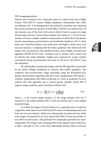
Chapter 4 number of counts and acquisition time, the whole body images reconstructed from split 50% count data served as surrogates for images acquired at 1.5min per bed position (Figure 4.1). Figure 4.1: Illustration of a PET image of a typical mCRPC patient with extensive [18F]FDHT-avid bone metastases reconstructed with (A) 100% counts EARL1, (B) 50% counts EARL1, (C) 100% counts EARL2, and (D) 50% counts EARL2. Axial (left column), coronal (middle column), and sagittal views (right column) are shown. 88 SUVbw SUVbw SUVbw SUVbw
