
Page 56 - 18F-FDG PET as biomarker in aggressive lymphoma; technical and clinical validation
P. 56
Chapter 3
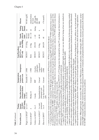
Table 2 Continued
checked before 90-160 mg/dL <200 mg/dL <7·8 mmol/L the exam (84-188) NR F-FDG uptake at some disease +- 60 fasting (min) (hours) Uptake Fasting inter val period 18 60 +- 10 ≥6 60 (n=6 ≥6 90 min) 60-90 6 60 ≥6 dose (MBq) F-FDG 37 /10 kg 370-555 240-259 2-5/kg 5·3/kg 18 F-FDG uptake at disease sites greater than mediastinum, but less than or equal to physiologic uptake in liver. Scan Procedures PET only PET/CT or PET/CT PET/CT PET/CT PET/CT Both = positive= according to a SUVmax measurement >2.5. Equivocal PET/CT or CT findings were then interpreted by using CT scan findings and clinical information to = a negative PET scan was defined as having no residual abnormal uptake or a minimal residual uptake. A positive scan was defined as having at least one residual site 3 experienced radiologists
= negative= no pathological tracer uptake was shown, unequivocally positive= areas of focal uptake localized to sites of previous disease (in this sense representing a residual disease or a disease relapse), within asymmetrical lymph nodes, or within lymph nodes unlikely to be affected by inflammation (mediastinal, except for hilar, and abdominal). = negative was defined as no evidence of disease. MRU was defined as low-grade uptake of FDG (just above background) in a focus within an area of previously noted 2 nuclear F-FDG uptake with intensity greater than liver at a site of known disease. Mixed response= reduction in readers
= 4 categories; Negative/CR= resolution of abnormal F-FDG uptake at sites of disease identified on staging PET with any residual F-FDG uptake less than or equal to disease reported by the nuclear medicine physicians. Positive was defined as increased uptake suspicious for malignant diseases, which did not have a benign explanation. 2 NM
1 NM
2 NM Interpretation
Deauville 4-5
Criteria for
positive scan
= results presented for central review results only, local review results are based on SUVmax lesion > SUVmax of mediastinal bloodpool. 18
b 18 18 associated with an intensity markedly superior to local background as previously described (Haioun Blood 2005). 3 or 4 Last day before new Custom
Days after previous
Sites of known physiological uptake that showed symmetrical uptake were not described in the report. 2 Median 14 Custom
treatment median
F-FDG uptake at other existing or new sites.
13 (4-27)
2 or 3 3 weeks IHP
(range)
treatment subsequent cycle
cycle
distinguish vascular/bowel activity from residual FDG uptake at the previously involved site.
= after 3 cycles R-CHOP21 or midtreatment in case of R-VNCOP-B or R-MACOP-B. Zinzani et al (2011) 3 or mid- Immediately before Custom
e
PET timing
18
Interim
F-FDG
Timing
2, 3 or 4
the mediastinal blood pool, CR-MRU= residual low-level
18
49
51
52 f
18
50 d
53 g
Positive=residual or increased
Pregno et al (2012)
Safar et al (2012)
Cashen et al (2011)
Zhao et al (2007)
sites, with increased
a
c
d
e
f
g
Abbreviations: MBq=megabecquerel, min=minutes, NR=not reported, NM=nuclear medicine physician, RAD=radiologist, IHP= international harmonization project criteria, IQR=interquartile range, SD= standard deviation, dept=department, (R-)CHOP= (rituximab,) cyclophosphamide, doxorubicin, vincristine, prednisone, R=rituximab, R-ACVBP= rituximab, doxorubicin, vindesine, bleomycin, prednisone.
54
Glucose
Interpreters
First author, year
