
Page 47 - Tyrosine-Based Bioconjugations - Jorick Bruins
P. 47
Mass spectrometry (MS) analysis of the SPOCQ reaction show a main product corresponding to the product resulting from oxidation and cycloaddition of 1 (Figure 2B, C), with a deviation of ±1 Da, Figure 2B displays the MS profile of LamA–G4Y prior to oxidation, in agreement with the calculated mass, whereas Figure 2C exhibits LamA–G4Y after oxidation and SPOCQ. The desired product was identified as a peak with molecular weight 32647 Da, indicating an increase of 880 Da (calculated 879 Da) from oxidation of the tyrosine and subsequent conjugation with 1. The oxidized LamA that underwent aspecific conjugation by a nucleophilic amino acid residue addition was also clearly detected on MS with a mass increase of approximately 13 Da. Further experiments showed that conjugation via SPOCQ could also be performed at 4 and 16 °C and ambient temperature with no observable difference in efficiency (Figure S2).
Trastuzumab. Having successfully demonstrated the suitability of SPOCQ for C-terminal protein conjugation, its usefulness for site-specific modification of monoclonal antibodies was investigated next. Trastuzumab with genetically engineered tetra-glycyltyrosine on both light chains (Tras[LC]G4Y) was transiently expressed in CHO-K1 and purified by protein A affinity chromatography. Next, Tras[LC]G4Y was subjected to identical conditions for conjugation with 1 (5 eq.) by SPOCQ at 16 °C as described for LamA. As expected, SDS-PAGE analysis indicated exclusive conversion of Tras[LC]G4Y upon incubation with both 1 and mTyr (SI Figure 1A), while no reaction was detected on either chain for native trastuzumab under identical conditions. Mass spectrometric analysis confirmed successful addition of 1 via SPOCQ (SI Figure 1 B-C). It is worth pointing out that the reaction also proceeded at 4 °C, albeit much slower, but to our surprise, no fluorescent protein could be detected at higher temperature (37 °C), possibly via competing intramolecular reaction of the intermediate quinone with nearby lysine or histidine side chains (Figure S4).
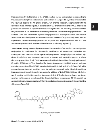
Figure 3. Chemical structure of BCN-MMAF 2.
Protein labelling via SPOCQ
45
2
