
Page 72 - Molecular features of low-grade developmental brain tumours
P. 72
3
CHAPTER 3
70
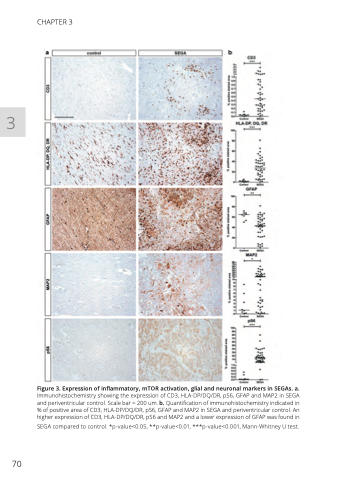
Figure 3. Expression of inflammatory, mTOR activation, glial and neuronal markers in SEGAs. a.
Immunohistochemistry showing the expression of CD3, HLA-DP/DQ/DR, pS6, GFAP and MAP2 in SEGA and periventricular control. Scale bar = 200 um. b. Quantification of immunohistochemistry indicated in % of positive area of CD3, HLA-DP/DQ/DR, pS6, GFAP and MAP2 in SEGA and periventricular control. An higher expression of CD3, HLA-DP/DQ/DR, pS6 and MAP2 and a lower expression of GFAP was found in
SEGA compared to control. *p-value<0.05, **p-value<0.01, ***p-value<0.001, Mann-Whitney U test.
