
Page 16 - Molecular features of low-grade developmental brain tumours
P. 16
1
CHAPTER 1
of the mTOR pathway (indicated by increased phosphorylation of S6K1 and S6) (figure 3G) 69,70,91. Recently, three distinct histological cortical tuber subtypes (designated as types A, B and C) have been described based on the proportion of calcifications, dysmorphic neurons and giant cells 80.
Cortical tubers can already develop during embryonic brain development and can therefore be detected prenatally 70,93-97. Additionally, elevated mTORC1 activation has been shown in prenatal cortical tubers, before seizure development 70,97,98. Seizures in the fetus are not physiologically possible before 24–25 weeks of gestation because synaptogenesis in the cortical plate is not initiated until 22 weeks of gestation 99. These findings indicate the importance of the mTOR pathway during brain development and highlight the role of mTOR dysregulation in pathological changes of the brain 69,70.
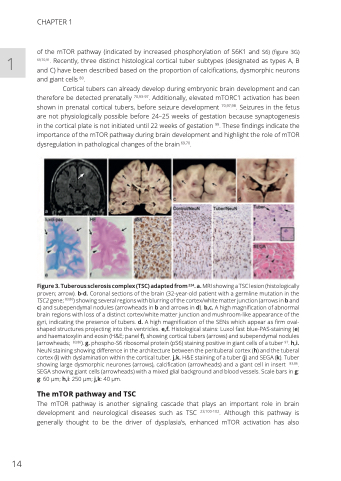
Figure 3. Tuberous sclerosis complex (TSC) adapted from 224. a. MRI showing a TSC lesion (histologically proven; arrow). b-d. Coronal sections of the brain (32-year-old patient with a germline mutation in the TSC2 gene; 83,86) showing several regions with blurring of the cortex/white matter junction (arrows in b and c) and subependymal nodules (arrowheads in b and arrows in d). b,c. A high magnification of abnormal brain regions with loss of a distinct cortex/white matter junction and mushroom-like appearance of the gyri, indicating the presence of tubers. d. A high magnification of the SENs which appear as firm oval- shaped structures projecting into the ventricles. e,f. Histological stains: Luxol fast blue-PAS-staining (e) and haematoxylin and eosin (H&E; panel f), showing cortical tubers (arrows) and subependymal nodules (arrowheads; 83,86). g. phospho-S6 ribosomal protein (pS6) staining positive in giant cells of a tuber 97. h,i. NeuN staining showing difference in the architecture between the perituberal cortex (h) and the tuberal cortex (i) with dyslamination within the cortical tuber. j,k. H&E staining of a tuber (j) and SEGA (k). Tuber showing large dysmorphic neurones (arrows), calcification (arrowheads) and a giant cell in insert 83,86. SEGA showing giant cells (arrowheads) with a mixed glial background and blood vessels. Scale bars in g: g: 60 μm; h,i: 250 μm; j,k: 40 μm.
The mTOR pathway and TSC
The mTOR pathway is another signaling cascade that plays an important role in brain development and neurological diseases such as TSC 23,100-102. Although this pathway is generally thought to be the driver of dysplasia’s, enhanced mTOR activation has also
14
