
Page 18 - The role of advanced echocardiography in patients with ischemic heart disease - Rachid Abou
P. 18
Chapter one. General introduction and outline of the thesis
practice, since 1) images obtained from the axial views for LV GLS measurement have better lateral resolution than short-axis images, 2) LV GLS is obtained from the entire length of the LV and therefore includes a greater amount of myocardial tissue, when compared to the short-axis views and 3) radial and circumferential strain demonstrate lower reproducibility than LV GLS.3
Figure 1. Schematic representation of the left ventricular myocardial wall. On the left, the left ventricular myocardial wall is formed by three layers: the subendocardial layer with the fibres arranged in the direction of a right-handed helix (hand and arrow), the subepicardial layer with the fibres oriented as a left-handed helix (hand and arrow)
and the mid-myocardial with the fibres oriented circumferentially. When the subendocardial and subepicardial layers shorten in opposite directions and the mid-myocardial layer shortens in the circumferential direction, the ventricle shortens in the longitudinal and circumferential direction and thickens in the radial direction (arrows).
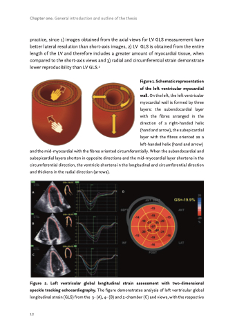
Figure 2. Left ventricular global longitudinal strain assessment with two-dimensional speckle tracking echocardiography. The figure demonstrates analysis of left ventricular global longitudinal strain (GLS) from the 3- (A), 4- (B) and 2-chamber (C) and views, with the respective
12
