
Page 114 - The role of advanced echocardiography in patients with ischemic heart disease - Rachid Abou
P. 114
Chapter six. Layer-specific LV GLS and prognosis
coronary angiography performed upon admission, the culprit lesion was identified and the final Thrombolysis In Myocardial Infarction (TIMI) flow after primary percutaneous coronary intervention was evaluated and registered. Multi-vessel disease was defined as the presence of more than one vessel with luminal narrowing ≥70%. Cardiovascular medications at hospital discharge were recorded and optimized at the discretion of the treating physician.
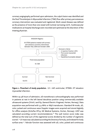
Figure 1. Flowchart of study population. LV = left ventricular; STEMI= ST elevation myocardial infarction.
Within 24-48 hours of admission, 2D-transthoracic echocardiography was performed in patients at rest in the left lateral decubitus position using commercially available ultrasound systems (Vivid 7 and E9; General Electric Vingmed, Horten, Norway). Data acquisition was performed with 3.5-MHz or M5S transducers. Standard M-mode, 2D, color, pulsed and continuous wave Doppler images were acquired and stored digitally for offline analysis (EchoPac BT13; GE Medical Systems, Horten, Norway). LVEF was calculated according current recommendations.10 The wall motion score index was defined as the total sum of the segmental scores divided by the number of segments scored .10 LV mass was calculated according the Devereux formula, and indexed for body surface area 10. Valvular function was assessed with 2D, color, pulsed and continuous
108
