
Page 102 - Strategies for non-invasive managementof high-grade cervical intraepithelial neoplasia - prognostic biomarkers and immunotherapy Margot Maria Koeneman
P. 102
Chapter 5
on hematoxylin and eosin staining (figure 1). Observational management of CIN 2 consisted of serial PAP smears after 6, 12, and 24 months. Patients were included in this study if they had at least one follow-up visit with cervical cytology during the first year. A new colposcopy was planned in case of PAP 3a2 (cytological HSIL) or higher at either of the follow-up visits. Exclusion criteria were previous treatments for high-grade CIN and concomitant CIN 3 or higher. For patients who completed the follow-up schedule, disease regression was defined as PAP 1 cytology at the 24 month follow-up or ≤CIN 1 histology at the 24 month follow-up and no diagnosis of >CIN 2 before the 24 month follow-up visit. For patients who did not complete the 24 month follow-up schedule, those with PAP 1 cytology at the last follow-up visit were included in the regression group, and all other patients were included in the persistence group.
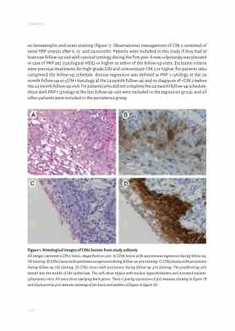
Figure 1. Histological images of CIN2 lesions from study subjects
All images represent a CIN 2 lesion, magnification 40x. A) CIN2 lesion with spontaneous regression during follow-up, HE staining. B) CIN2 lesion with spontaneous regression during follow-up, p16 staining. C) CIN2 lesions with persistence during follow-up, HE staining. D) CIN2 lesion with persistence during follow-up, p16 staining. The proliferating cells extend into the middle of the epithelium. The cells show atypia with nuclear hyperchromasia and increased nuclear- cytoplasmic ratio. All cases show overlying koilocytosis. There is patchy expression of p16 immuno staining in figure 1B and block positive p16 immuno staining of the basal and middle cell layers in figure 1D.
100
